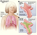
Αρχείο:Bronchitis.jpg
Bronchitis.jpg (475 × 445 εικονοστοιχεία, μέγεθος αρχείου: 100 KB, τύπος MIME: image/jpeg)
Ιστορικό αρχείου
Κλικάρετε σε μια ημερομηνία/ώρα για να δείτε το αρχείο όπως εμφανιζόταν εκείνη τη στιγμή.
| Ώρα/Ημερομ. | Μικρογραφία | Διαστάσεις | Χρήστης | Σχόλια | |
|---|---|---|---|---|---|
| τελευταία | 15:19, 12 Νοεμβρίου 2013 |  | 475 × 445 (100 KB) | CFCF | User created page with UploadWizard |
Συνδέσεις αρχείου
Τα παρακάτω λήμματα συνδέουν σε αυτό το αρχείο:
Καθολική χρήση αρχείου
Τα ακόλουθα άλλα wiki χρησιμοποιούν αυτό το αρχείο:
- Χρήση σε af.wikipedia.org
- Χρήση σε ar.wikipedia.org
- Χρήση σε bg.wikipedia.org
- Χρήση σε bn.wikipedia.org
- Χρήση σε en.wikipedia.org
- Χρήση σε et.wikipedia.org
- Χρήση σε eu.wikipedia.org
- Χρήση σε fa.wikipedia.org
- Χρήση σε fi.wikipedia.org
- Χρήση σε fr.wikipedia.org
- Χρήση σε ga.wikipedia.org
- Χρήση σε hr.wikipedia.org
- Χρήση σε hy.wikipedia.org
- Χρήση σε hyw.wikipedia.org
- Χρήση σε id.wikipedia.org
- Χρήση σε io.wikipedia.org
- Χρήση σε is.wikipedia.org
- Χρήση σε it.wikibooks.org
- Χρήση σε it.wikiversity.org
- Χρήση σε ja.wikipedia.org
- Χρήση σε ko.wikipedia.org
- Χρήση σε mk.wikipedia.org
- Χρήση σε or.wikipedia.org
- Χρήση σε pt.wikipedia.org
- Χρήση σε ro.wikipedia.org
- Χρήση σε simple.wikipedia.org
- Χρήση σε sq.wikipedia.org
- Χρήση σε sr.wikipedia.org
- Χρήση σε sv.wikipedia.org
- Χρήση σε th.wikipedia.org
- Χρήση σε tr.wikipedia.org
- Χρήση σε uk.wikipedia.org
- Χρήση σε vi.wikipedia.org
- Χρήση σε www.wikidata.org
- Χρήση σε zh-yue.wikipedia.org
- Χρήση σε zh.wikipedia.org


